In contrast to the conventional chiral crystalline materials formed by solids with chiral space groups, chiral mesostructured inorganic crystals can be inductively assembled by inorganic units with achiral space groups. Due to coiled crystal lattice and helical stacking, they exhibit unique physical and chemical chiral anisotropy. However, it is very difficult to analyze such structures. Spectroscopy and X-ray diffraction only provide overall structural information. Although scanning electron microscopy can help people observe the chiral morphology of twisted or helically stacked nanocrystals, it is unlikely to determine the orientation of small-angle helically stacked nanocrystals. However, precession electron diffraction, electron backscattering diffraction, nanobeam diffraction, and other technologies can resolve the chiral crystals formed by chiral space groups. Still, they are not suitable for forming chiral mesoscopic inorganic crystals by non-chiral space groups. At present, it is often necessary to manually tilt the goniometer to make the crystal belt axis of different parts of the crystal align with the positron beam to calculate the corresponding angular deflection and pitch to determine the torsional chirality of chiral inorganic crystals. However, it is still challenging to determine the synchronization of graded chirality and quantify and analyze the structural information such as specific torsional angle, rotational axis, and stacking.
Recently, Professor HAN Lu's research team from the School of Chemical Science and Engineering of our university proposed a general approach to determine chiral hierarchical mesostructures based on three-dimensional electron diffraction tomography (3D EDT). This approach reconstructs a complete set of three-dimensional electron diffraction data sets from two-dimensional electron diffraction patterns within the tilt range of the transmission electron microscope goniometer. The corresponding relationship between the electron diffraction pattern based on reciprocal space modulation and the crystal structure of mesoscopic materials in positive space, and the corresponding diffraction data processing and calculation methods are used to synchronously solve the multi-level chiral structure information of inorganic crystals. Their research findings were published in Nature Communications under "Synchronous quantitative analysis of chiral mesostructured inorganic crystals by 3D electron diffusion tomography".
HAN Lu's research team at the School of Chemical Science and Engineering proposed a general approach for quantitative analysis of chiral mesostructured inorganic crystals by three-dimensional electron diffraction tomography. Their research findings were published in Nature Communications.
First, the researchers built five representative crystal structures and established the correspondence between the positive space modulation structure and the intensity distribution and relative position change of diffraction spots in the reciprocal space through the Fourier transform. The reciprocal space of a single crystal has independent diffraction with the periodic arrangement; The diffraction plane of the chiral mesoscopic structure changes with the bending of the crystal structure. The intensity distribution of the diffraction points shows a "bending profile.” The diffraction has a central symmetric arch intensity distribution consistent with the bending angle and direction of the crystal and shows helical characteristics.
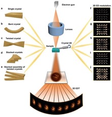
Five representative crystal structures
The helically stacked nanocrystals present a three-dimensional electron diffraction data set formed by superpositioning multiple diffraction patterns. Therefore, the first-level chiral twist of the mesoscopic crystal structure can be revealed by the intensity distribution shape of the diffraction spots in the three-dimensional reciprocal space lattice. In contrast, the correspondence between the main diffraction spots and the surrounding satellite diffraction spots represents the second-level helical chirality of the nanocrystal stack. According to the change of the diffraction spots and the corresponding sample measurement length, the twist direction, pitch, hierarchical chirality, and other information of the chiral materials can be accurately calculated. Although it is difficult to retrieve this chiral mesostructured information by conventional electron microscopy, they are well preserved in the reconstructed three-dimensional reciprocal space.
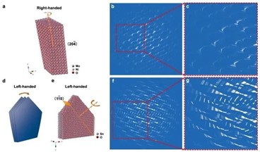
The method has been applied to the hydrothermal synthesis of chiral mesoscopic nickel molybdate (L/D-CNM) with chiral molecule L/D-threonine as a symmetry-breaking agent and structure-directing agent, Fe2SO4 • 7H2O as an inorganic precursor. Through the periodic arc diffraction of three-dimensional electron diffraction and the helical arrangement of diffraction in the vertical direction, the first level chirality of D-CNM lattice torsion has been successfully resolved, revealing that the central axis of D-CNM rotation is in the (20-4) crystal plane normal direction. The D-CNM twist angle of 25.1 ° and the helix of about 33.0 ° μ m are determined by the average central angle of arc diffraction perpendicular to the central axis of rotation of the three-dimensional electron diffraction pattern at both ends of the D-CNM rod particle.
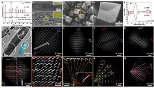
The method is applied to the chiral molecule L/D-threonine as a symmetry-breaking agent and structure-directing agent
At the same time, the authors also revealed the primary lattice torsion chirality and secondary helix stacking chirality of chiral mesoscopic tin dioxide crystal (L-CTD) induced by ammonium L-tartrate. The axis of the L-CTD rotation center is (1-10) the normal direction of the crystal plane, the torsion angle is 2.2 °, and the helix is about 26.2 μ m。 Through a series of diffraction spots superposed around the origin in the slice electron diffraction of the three-dimensional electron diffraction data set, it is found that there is a second-level chirality of the helical stacking of nanosheets in the L-CTD nanoplate. According to the average rotation angle of the diffraction, the rotation dihedral angle of the nanosheet is 1.3 °.
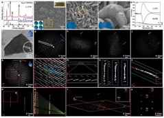
The method also reveals the primary lattice torsion chirality and secondary helix stacking chirality of chiral mesoscopic tin dioxide crystal (L-CTD) induced by ammonium L-tartrate
Finally, the authors verified the precision and effectiveness of the three-dimensional electron diffraction reconstruction method in judging multi-level chiral self-assembly through calculation and simulation. This study developed the application of three-dimensional electron diffraction quantitative analysis in the structural analysis of different chiral mesoscopic crystal materials. It provided an insight into the understanding of structural–chiral anisotropy at the fundamental level and the construction and structure research of chiral inorganic materials in the future. In addition, this structural relationship and analytical method can be extended to a variety of curved crystals, defective crystals, and self-assembled structures stacked with nanocrystals, and has important research value for chiral science, nanomaterial chemistry, and crystallography.
Three-dimensional electron diffraction reconstruction approach
The corresponding author of this paper is Professor Han Lu from the School of Chemical Science and Engineering, and the first author is Ai Jing, a doctoral student from the School of Chemical Science and Engineering. The research team got help from Professor CHE Shunai's team, Professor Osamu Terasaki of Shanghai University of Science and Technology, and Professor Peter Oleynikov's team. The research was supported by the National Natural Science Foundation of China, the Special Fund for Basic Scientific Research, the National Key Research and Development Plan, the Science Fund of Shanghai Municipal Science and Technology Commission, and the Research Center of Shanghai University of Science and Technology for High-resolution Electron Microscopy.
Paper link: https://www.nature.com/articles/s41467-018-06539-w